← rhabdomyosarcoma mri cervial Rhabdomyosarcoma embryonal radiology radiopaedia leiomyoma uterus lbael diagram 209 best uterus images on pinterest →
If you are looking for MRI - FOOT you've came to the right place. We have 35 Pics about MRI - FOOT like MR image quality - Radiology Cafe, MRI SCAN - Fitter Feet For LifeFitter Feet For Life and also MRI - FOOT. Read more:
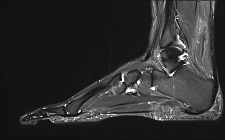
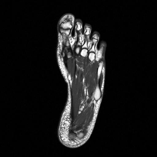
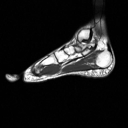
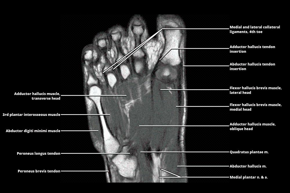
MRI - FOOT
 www.wangmd.com
mri
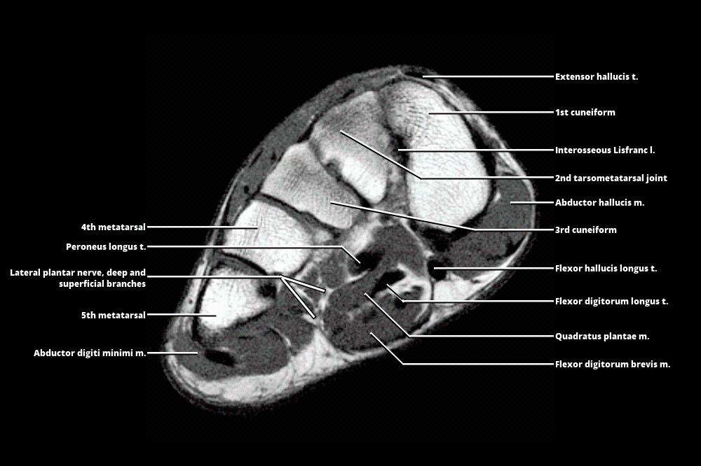
MRI Of The Fore-foot; Short-axis (a) T2 FS, Sagittal (b) Non-contrast
 www.researchgate.net
axis mri fore t2 imaging spectrum findings diabetic t1w
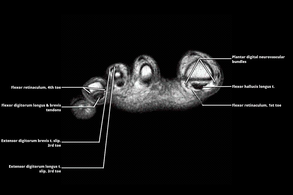
(a–e) MRI Examination Of The Foot Sagittal (a–d) And Axial (e) T1 WI
 www.researchgate.net
foot mri examination axial sagittal wi
MRI - FOOT
 www.wangmd.com
mri
Foot MRI Scan | Medserena Open MRI
 www.medserena.co.uk
mri scan axial
Anatomy Of The Foot And Ankle - MRI | E-Anatomy
 www.imaios.com
MRI Of The Foot • APPLIED RADIOLOGY
 appliedradiology.com
mri foot ali2
Normal Magnetic Resonance Imaging Anatomy Of The Ankle & Foot
 www.mri.theclinics.com
mri foot ankle anatomy cuboid normal ligament lateral calcaneocuboid calcaneal joint magnetic resonance imaging figure tuberosity large
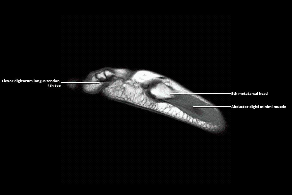
Foot Muscles Mri Anatomy - MRI Of The Ankle: Detailed Anatomy - W
 fredrickson-nortonsamrawit.blogspot.com
mri radiology
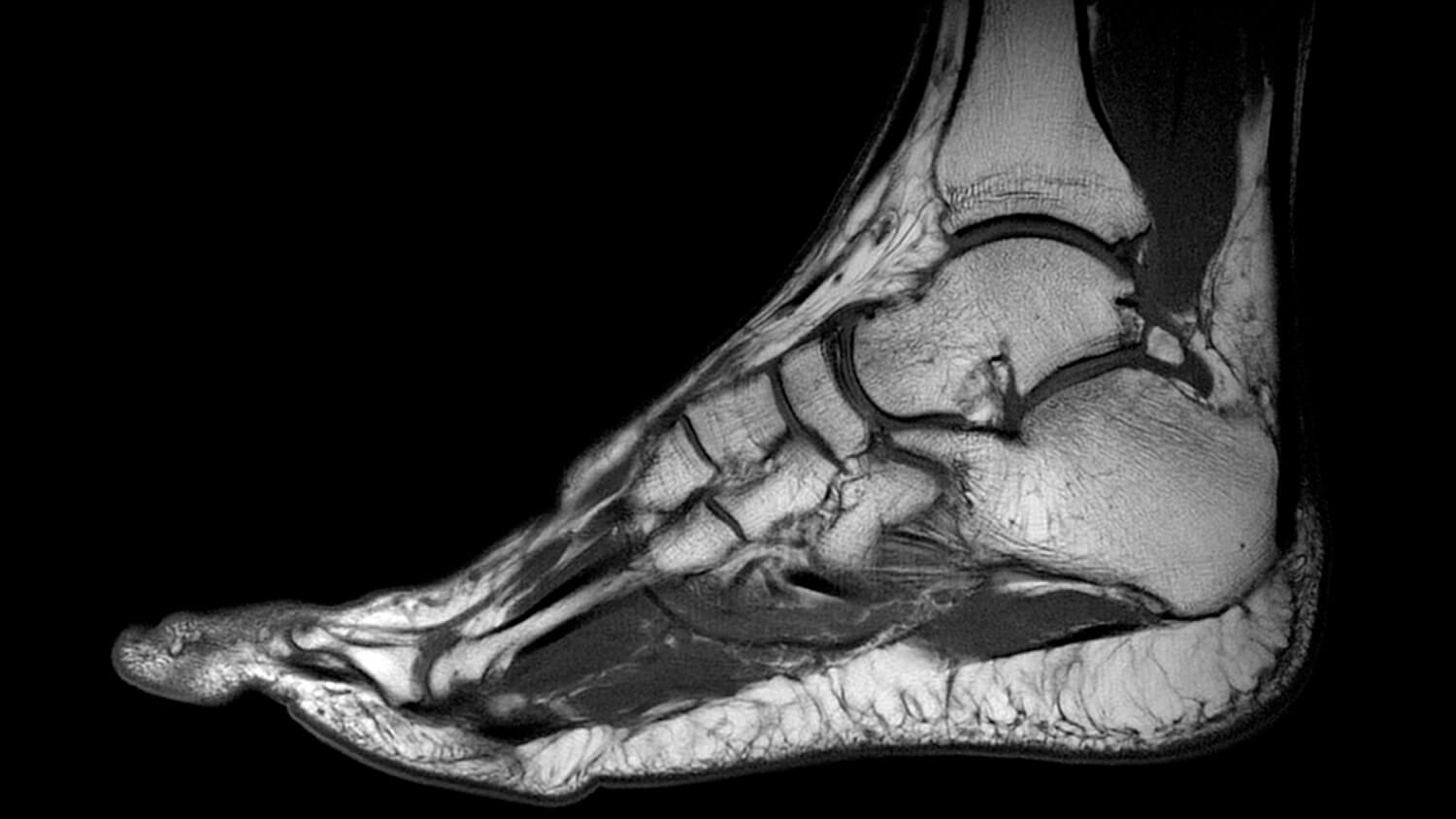
Normal Ankle MRI | Radiology Case | Radiopaedia.org
 radiopaedia.org
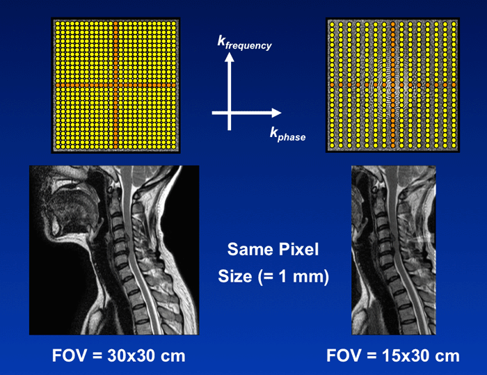
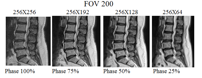
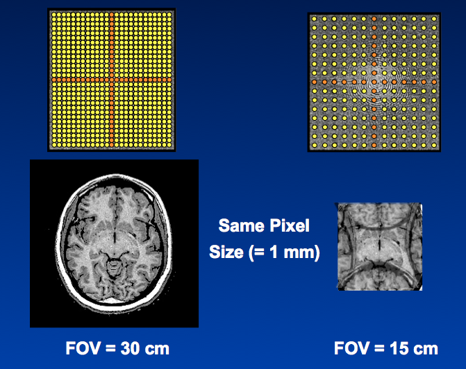
Rectangular FOV - Questions And Answers In MRI
 www.mriquestions.com
Normal Left Foot MRI | Radiology Case | Radiopaedia.org
 radiopaedia.org
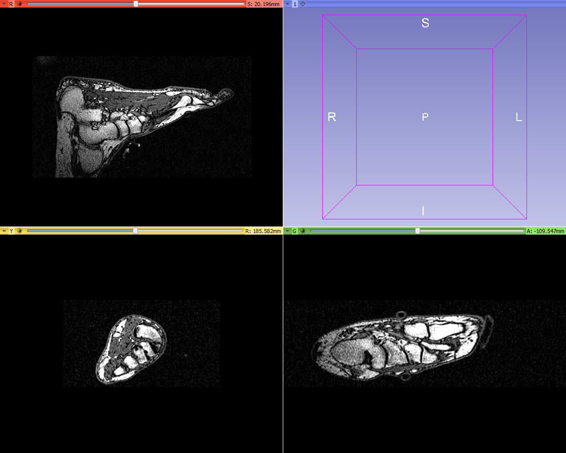
Segmentation Of A Foot MRI Scan - Mike At Medical Models - Embodi3D.com
 www.embodi3d.com
mri foot scan segmentation embodi3d medical aged middle using woman am available here data
Normal Foot MRI | Image | Radiopaedia.org
 radiopaedia.org
MRI Of The Foot • APPLIED RADIOLOGY
 appliedradiology.com
mri foot ali2
Feet MRI
 centres.exeter.ac.uk
mri foot right feet coronal ms centres exeter ac
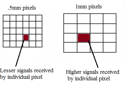
MRI Resolution And Image Quality | How To Manipulate Mri Scan Parameters
 mrimaster.com
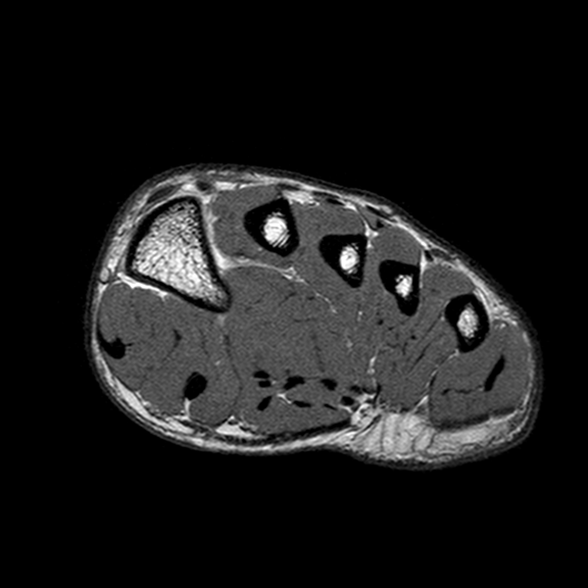
MRI Of The Right Feet: Axial T1-weighted Image Showing Nodular
 www.researchgate.net
mri axial curvilinea cutanea piede margine mediale weighted
Feet MRI
 centres.exeter.ac.uk
feet ffe centres exeter ac
MRI Imaging Techniques: MRI Technologist Role In Foot/Ankle Positioning
 imaging-mri.blogspot.com
positioning mri ankle anatomy imaging foot gastrocnemius achilles insertion must cover technologist role
MRI - FOOT
 www.wangmd.com
mri
MRI - FOOT
 www.wangmd.com
mri atlas
MRI Scan For Ankle Injury Melbourne - Melbourne Radiology
 www.melbourneradiology.com.au
ankle mri tendon achilles fasciitis tear plantar radiology ultrasound
Normal Magnetic Resonance Imaging Anatomy Of The Ankle & Foot
 www.mri.theclinics.com
ankle anatomy foot mri normal coronal resonance imaging magnetic t1 fig
Foot Muscles Mri - MRI With User Outlined Plantar Intrinsic And
 blackbirdsewing.blogspot.com
mri foot plantar anatomy muscles intrinsic muscle toes outlined theclinics aponeurosis tendon imaging extrinsic minimi digiti
MR Image Quality - Radiology Cafe
 www.radiologycafe.com
MRI Of The Foot | Radiology Key
 radiologykey.com
mri foot radiology
MRI - FOOT
 www.wangmd.com
mri
[Figure, T2-weighted MRI Image Of A...] - StatPearls - NCBI Bookshelf
![[Figure, T2-weighted MRI image of a...] - StatPearls - NCBI Bookshelf](https://www.ncbi.nlm.nih.gov/books/NBK532250/bin/0126MR180002962_STIR__Long__Axis_18.jpg) www.ncbi.nlm.nih.gov
www.ncbi.nlm.nih.gov
MRI Resolution And Image Quality | How To Manipulate Mri Scan Parameters
 mrimaster.com
Disease Activity Evident On Foot MRI During Clinical Remission In
 www.rheumatologyadvisor.com
mri foot clinical remission arthritis rheumatoid evident rheumatologyadvisor
MRI - FOOT
 www.wangmd.com
mri atlas
Image | Radiopaedia.org
 radiopaedia.org
mri radiopaedia anatomy radiology pd muscle axial muscles imaging
MRI SCAN - Fitter Feet For LifeFitter Feet For Life
 fitterfeet.co.uk
mri scan foot feet does magnetic quality high work imaging
K-space: FOV - Questions And Answers In MRI
 www.mri-q.com
fov mri field space spacing
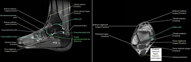
Ankle anatomy foot mri normal coronal resonance imaging magnetic t1 fig. Mri foot radiology. Mri of the fore-foot; short-axis (a) t2 fs, sagittal (b) non-contrast