← rhabdomyosarcoma mri axial thigh Rhabdomyosarcoma mri leiomyoma of uterus ct scan Ovarian mucinous cystadenoma and uterine leiomyoma – radiology cases →
If you are looking for Magnetic Resonance Imaging: Marrow Edema Patterns in Chronic Foot Pain you've visit to the right page. We have 35 Pictures about Magnetic Resonance Imaging: Marrow Edema Patterns in Chronic Foot Pain like Bone Marrow Edema Foot Treatment Quotes Update Viral - vrogue.co, Figure 1 from Bone marrow oedema syndrome of the foot and ankle in a and also Ankle Subchondroplasty - Foot Doctor San Diego / La Jolla Podiatrist. Read more:
Magnetic Resonance Imaging: Marrow Edema Patterns In Chronic Foot Pain
 mss-ijmsr.com
edema marrow foot magnetic imaging chronic resonance pain patterns
Figure 1 From Bone Marrow Oedema Syndrome Of The Foot And Ankle In A
 www.semanticscholar.org
Figure 1 From Painful Bone Marrow Edema Syndrome Of The Foot And Ankle
 www.semanticscholar.org
BONE MARROW EDEMA TREATMENTS - YouTube
 www.youtube.com
edema marrow
Bone Marrow Edema Is The Most Specific Finding For Rheumatoid Arthritis
 www.jrheum.org
edema bone marrow arthritis rheumatoid jrheum ra
MRI - FOOT
 www.wangmd.com
mri
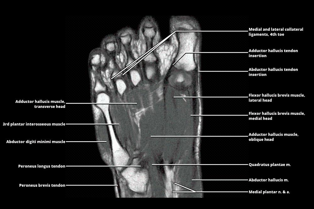
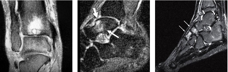
25: Magnetic Resonance Imaging Of Foot And Ankle Pathology
 musculoskeletalkey.com
foot ankle bone pathology marrow edema moderate coronal saturation intensity represents t2 signal fat figure
Regional Migratory Osteoporosis Complicated By Surgical Treatment: Case
 www.oatext.com
regional osteoporosis migratory bone edema talus showed imaging resonance magnetic marrow ankle foot figure which mri
Lateral Radiograph Of Ankles Showing Bony Enlargement At The
 www.researchgate.net
bony radiograph lateral ankles enlargement posterosuperior mri calcaneus commonly haglund mysterious syndrome aspect spurring
Be Aware Of Bone Marrow Edema Syndrome In Ankle Arthroscopy: A Case
 www.arthroscopyjournal.org
ankle edema marrow bone syndrome arthroscopy aware treated iloprost successfully
Bone Marrow Edema Patterns In The Ankle And Hindfoot: Distinguishing
 www.ajronline.org
Magnetic Resonance Imaging: Marrow Edema Patterns In Chronic Foot Pain
 mss-ijmsr.com
edema marrow foot imaging chronic resonance magnetic pain patterns close
25: Magnetic Resonance Imaging Of Foot And Ankle Pathology
 musculoskeletalkey.com
foot ankle pathology resonance imaging magnetic
Left Hip MRI Showing A Significant Reduction Of Bone Edema At Proximal
 www.researchgate.net
mri hip proximal significant edema femur
T2-weighted Fat Suppressed MRI Of The Right Foot; Bone Marrow Edema At
 www.researchgate.net
marrow weighted mri suppressed edema diffuse ankle proximal
MRI PD Image Showed Bone Marrow Edema With High Signal Intensity In The
 www.researchgate.net
marrow mri edema tibial intensity diaphysis
Same Foot As In Figure 3. Follow-up MRI Sagittal STIRsequence Shows
 www.researchgate.net
mri patchy sagittal edema marrow
Bone Marrow Edema Foot Treatment Quotes Update Viral - Vrogue.co
 www.vrogue.co
Mri 3 Months Later Showing Significant Regression Of The Right Hip
 www.researchgate.net
mri edema marrow femoral regression significant months regional
Figure 3 From Bone Marrow Edema Patterns In The Ankle And Hindfoot
 www.semanticscholar.org
Bone Marrow Edema - San Diego Running & Sports Injury Clinic
 www.sdri.net
edema marrow mri
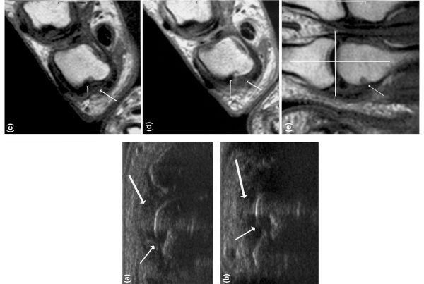
Figure 2 From Bone Marrow Edema Syndrome In The Foot And Ankle
 www.semanticscholar.org
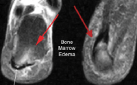
Bone Marrow Edema: Understanding What It Is - Orthopaedic Specialists
 louisvillebones.com
marrow bone edema understanding spongy specialists orthopaedic inside
Coloured MRI Scan Of Ankle Bones In The Human Foot - Stock Image - P116
 www.sciencephoto.com
mri ankle scan bones coloured simon fraser
Ankle Subchondroplasty - Foot Doctor San Diego / La Jolla Podiatrist
 www.footdocsandiego.com
ankle marrow edema bone foot mri
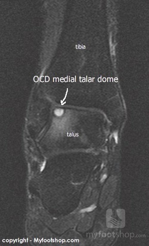
Bone Edema On MRI | MyFootShop.com
 www.myfootshop.com
bone edema myfootshop mri
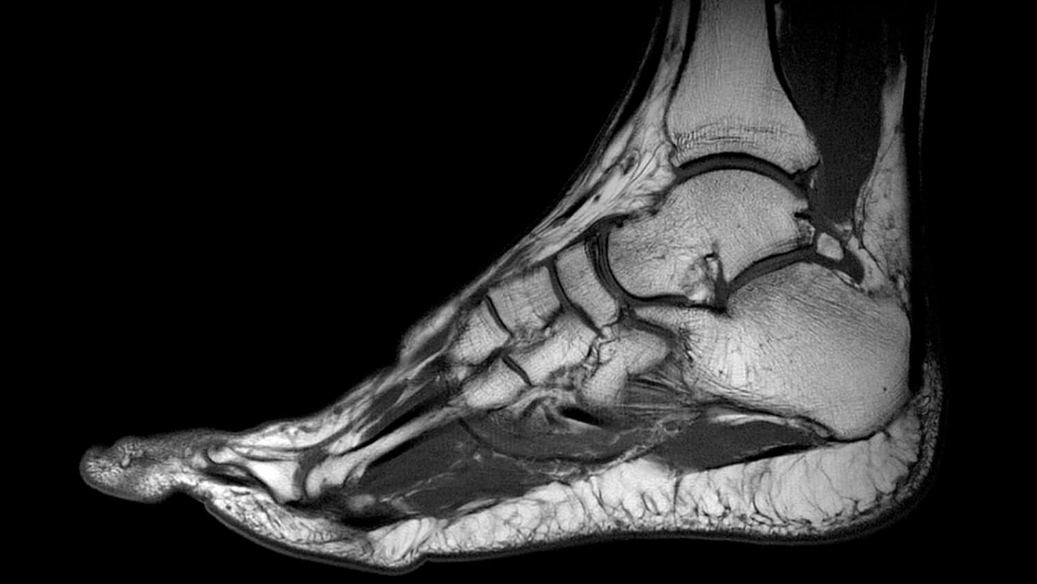
MRI SCAN - Fitter Feet For LifeFitter Feet For Life
 fitterfeet.co.uk
mri scan foot feet does magnetic quality high work imaging
Bone Edema On MRI May Predict Rheumatoid Arthritis | Radiology Report
 www.radiologyreport.org
mri bone edema predict may arthritis rheumatoid
Cureus | Effectiveness Of Iloprost In The Treatment Of Bone Marrow Edema
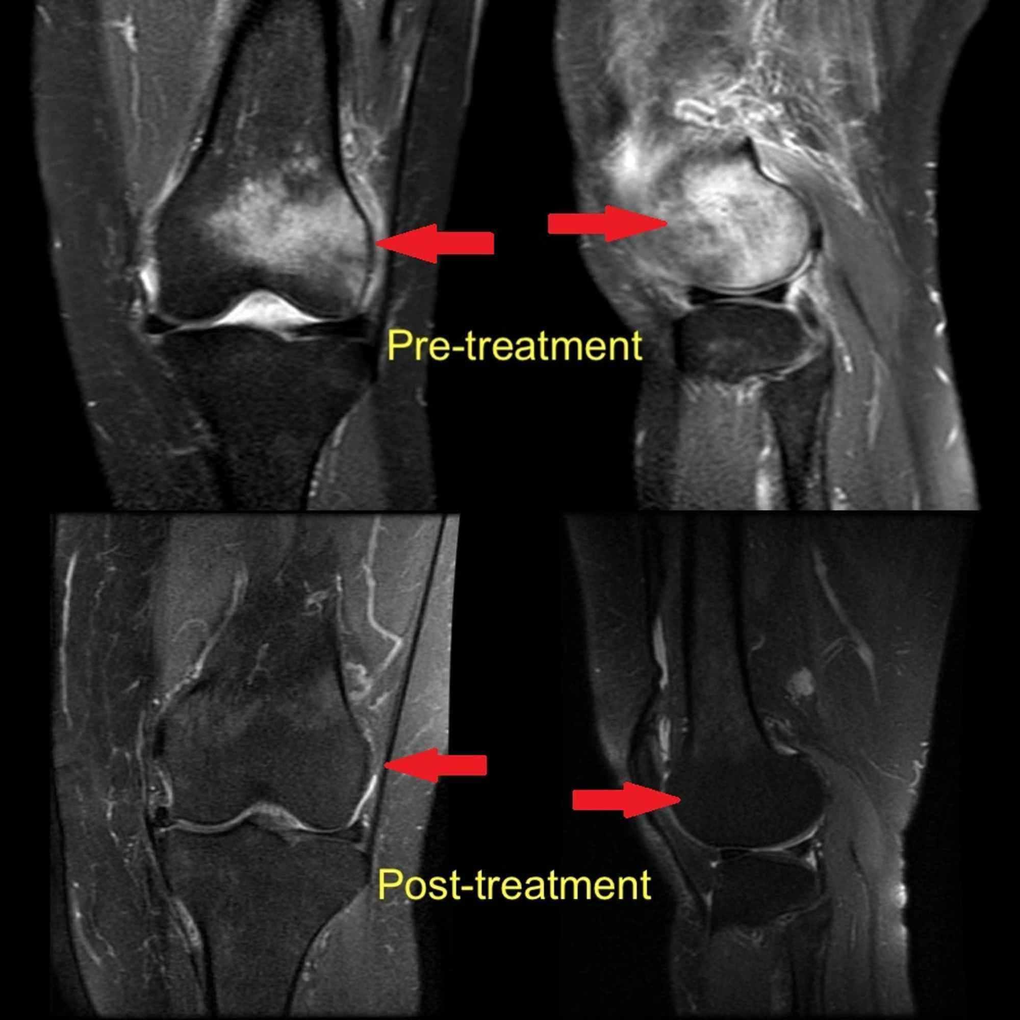 www.cureus.com
edema marrow mri femoral condyle lateral effectiveness iloprost cureus
Bone Marrow Edema Foot Treatment Quotes Update Viral - Vrogue.co
 www.vrogue.co
MRI Of Right Hip (a) Of 17 Year Old Female Shows Edema | Open-i
 openi.nlm.nih.gov
edema mri hip open right bone policy copyright access
Same Foot As In Figure 1. MRI Sagittal STIR-sequence Sh | Open-i
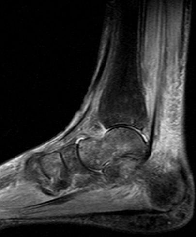 openi.nlm.nih.gov
mri foot bone crps edema figure marrow stir open sagittal same talus distal tibia diffuse subcutaneous tissue sequence skin openi
Bone Marrow Edema Pattern (BME) On MRI (low-signal Changes On
 www.researchgate.net
edema bone mri marrow signal bme weighted stir
Magnetic Resonance Imaging Foot Scan Showing Bone Marrow Edema At
 www.researchgate.net
Magnetic Resonance Imaging (MRI) Of Diabetic Foot Ulcers Shows Bone
 www.researchgate.net
Ankle edema marrow bone syndrome arthroscopy aware treated iloprost successfully. Marrow mri edema tibial intensity diaphysis. Same foot as in figure 1. mri sagittal stir-sequence sh